Patient Positioning
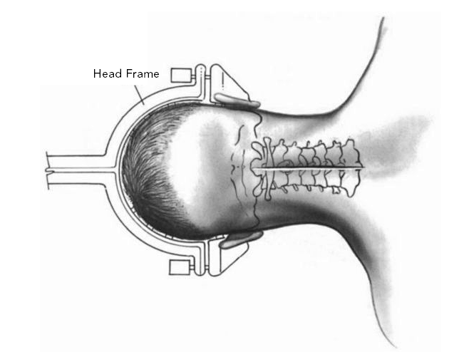
The patient is placed in a prone position with the head securely fixed, preferably in a slight flexed position. Moderate flexion of the neck reduces the overlap of facet joints and laminae, making the laminoplasty procedure more convenient and aiding in minimizing extradural and perivertebral venous bleeding.
Surgical Exposure
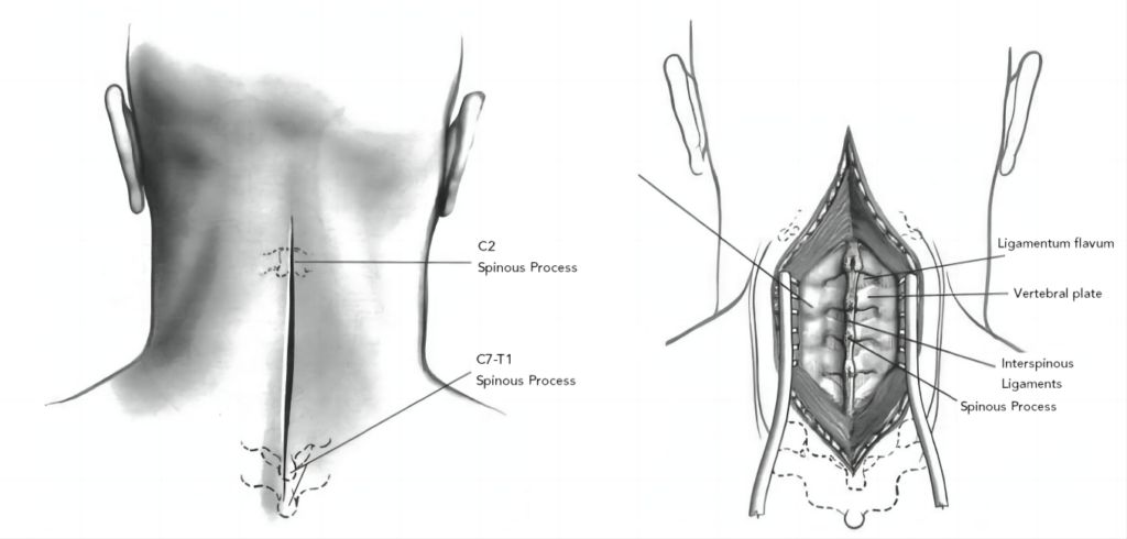
Taking C3 to C7 as an example, a midline posterior incision is made, exposing the area from the lower border of C2 to the upper border of T1. Dissection is performed bilaterally subperiosteally to the midportion of the cervical lateral masses, while muscles on the outer side are generally not dissected to reduce intraoperative bleeding. A slight separation of the extensor muscle attachment point at the lower border of C2 lamina exposes the C2-C3 lamina gap. Note: In cases where decompression is necessary up to the level of the pivot segment, preserving the intact pivot arch and most muscle attachments above it can also achieve decompression. Gun-type forceps and a burr are used to remove the lower border and middle cancellous bone and the anterior cortical bone of the C2 lamina, forming a dome-like shape.
Correct Door Opening
1.Door Opening and Axial Orientation
- If the spinal canal occupancy is symmetrical, the surgeon opens the door based on their preference.
- If the spinal canal occupancy is asymmetrical, the larger side is chosen for door opening to minimize disturbance to the spinal canal contents and reduce the risk of spinal cord injury.
- When simultaneous laminotomy decompression is required, the door opening should be on the same side as the laminotomy.
- Door opening instruments of choice are gun-type forceps and a burr.
2.Door Opening and Axial Position:
The door opening is placed at the junction of the lamina and lateral mass, aligning the outer edge of the bone trough with the inner edge of the pedicle.
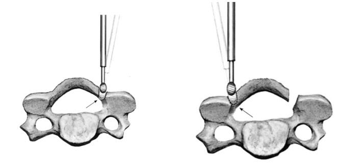
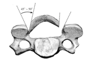
3. Door Opening and Axial Shape:
The door opening is performed in a vertical bone trough shape, creating a wedge-shaped bone trough with an angle of 45 degrees to 50 degrees, preserving 1mm thickness of cancellous and cortical bone.
4. Range of Door Opening:
The door opening includes all or part of the lamina of the spinal level above and below the target segment.
5.Grooving the Hinge Side:
After preparing the groove on the hinge side, the yellow ligament above and below the door area is cut, and the dura mater and veins are separated. The yellow ligaments at C2-C3 and C7-T1 are excised using forceps. Gentle flipping of the lamina while using a nerve dissector separates the veins, sequentially opening each lamina. Before opening the lamina, a curved probe is used to check for complete separation of the tissue below the lamina.
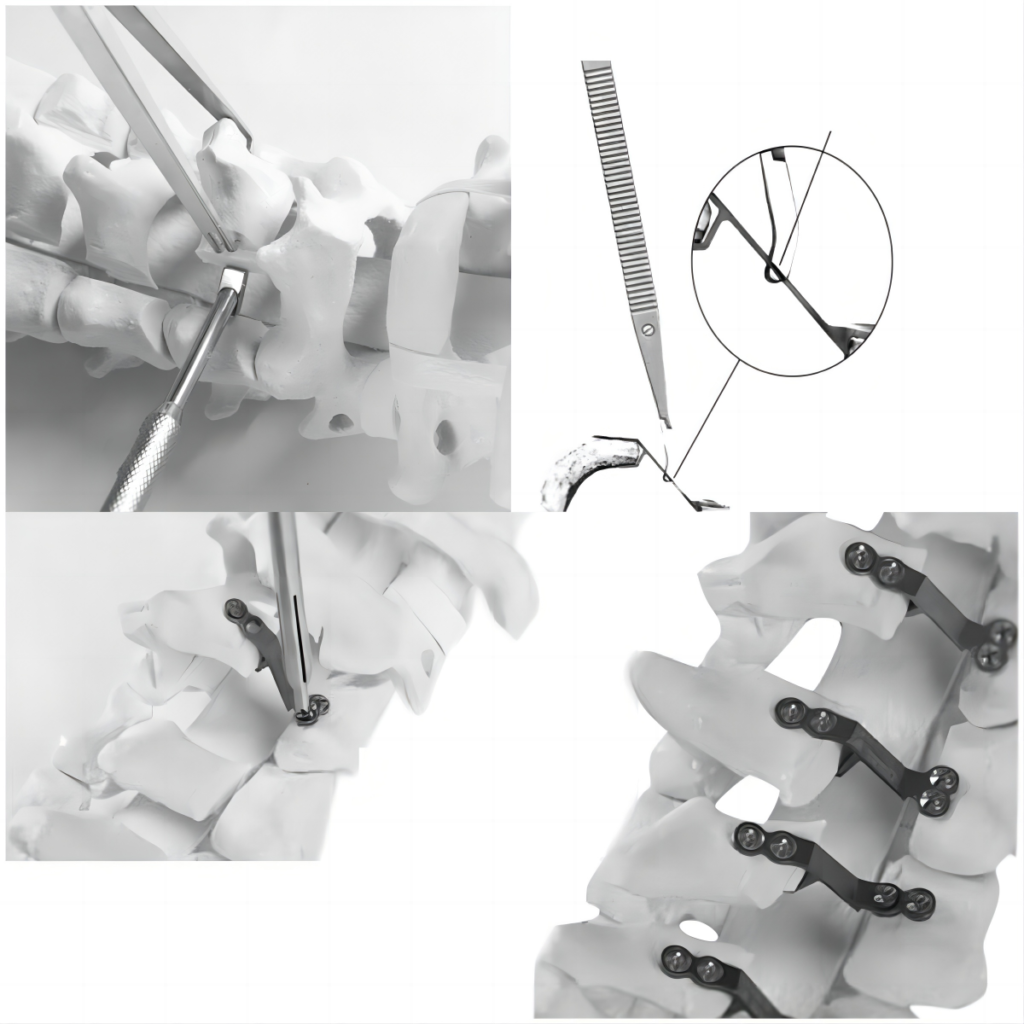
6.Door Opening Instruments:
On the door side, a 3mm burr is used to remove the outer cortical bone, some cancellous bone, and inner cortical bone. Then, a 1mm gun-type forceps is used to remove the remaining inner plate bone. On the hinge side, a 3mm burr is used to remove the outer cortical bone and some cancellous bone.
7.Range of Door Opening:
The door opening includes all or part of the lamina of the spinal level above and below the target segment.
8.Grooving the Hinge Side:
After preparing the groove on the hinge side, the yellow ligament above and below the door area is cut, and the dura mater and veins are separated. The yellow ligaments at C2-C3 and C7-T1 are excised using forceps. Gentle flipping of the lamina while using a nerve dissector separates the veins, sequentially opening each lamina. Before opening the lamina, a curved probe is used to check for complete separation of the tissue below the lamina.
Prevention of Door Closure and Collapse
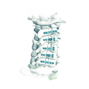
The positioning of the cervical plate system is determined using palpation to determine the appropriate model.
Manual or powered drills are used to create screw holes on the lateral mass. Screws are then implanted using a screwdriver.
Learn our product